Using our advanced imaging equipment, our Imaging and Image Analysis Unit facilitates and analyses scans for clinical services, trials, research as well as training researchers and clinicians.
Equipment
Dual-emission X-ray Absorptiometry (DXA)
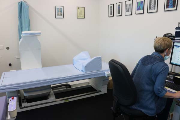
At our institute, we use Hologic – Horizon DXA machine to perform bone densitometry and body composition analysis. The usual scan areas for bone densitometry are lumbar spine, left and right hips, left and right forearms, and whole body.
Moreover, we perform body composition analysis which provides information about body fat and lean mass. At AIMSS, we use both National Health and Nutrition Examination Survey (NHANES) ranges and AIMSS ranges for the Australian population to report the body composition results.
Peripheral Quantitative Computed Tomography (pQCT)
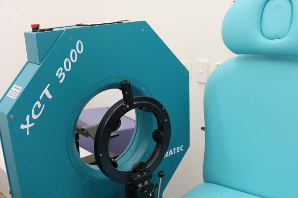
Using pQCT technology, we can extract quantitative information from bone, muscle, and fat in arms and legs. We use Stratec Medizintechnik – XCT 3000 machine at our institute to perform pQCT scans. The usual scan areas are 4% and 66% of tibia and radius. At AIMSS, we also perform pQCT scans for femur at 4% and 50% (midthigh).
Image Analysis – Tissue Compass™.
The Medical Imaging and Image Analysis department of AIMSS are developing a series of novel medical image analysis software known as Tissue Compass™.
Currently, the first version of this software is developed, which brings together advanced semi-automatic segmentation and automatic quantification techniques to help researchers analyse medical images in musculoskeletal science.
Tissue Compass™ 1.0 is validated against commercially available manual software and showed superior capabilities. Future versions of Tissue Compass™ will include novel Artificial Intelligence (AI) and Machine Learning technologies to fully automate segmentation and quantification tasks and provide prognostic and diagnostic assistant in research and clinical settings.
See the latest article reporting on the Validation of a Semiautomatic Image Analysis Software for the Quantification of Musculoskeletal Tissues.
If you are interested in receiving further information regarding Tissue Compass 1.0 or future versions complete a Tissue Compass™ Access Request.
Location
The Imaging and Image Analysis Unit is located at:
Level 4 WCHRE Building
Sunshine Hospital
176 Furlong Road
St Albans VIC 3021
General enquiries
This facility is used for clinical, research and training purposes. Access to imaging equipment is managed by the Imaging and Image Analysis unit.
Contact Mahdi Imani for more information.